POSTERIOR ANKLE IMPINGEMENT
"Painful Os Trigonum"
By: Robert H. Sheinberg, D.P.M., D.A.B.F.A.S., F.A.C.F.A.S.
Pain in the back of the ankle is very common in athletes in sports which require them to do a lot of jumping or in when they are on point or with the foot in a demi-point position.
CAUSES:
- Repetitive ankle trauma with overuse.
- There is an extra bone in the back of the ankle that fails to fuse to the main bone. It is called the "os trigonum". The os trigonum is connected to the main bone called the talus by a piece of cartilage. It is called a synchondrosis. With repetitive motion of the foot in the down position the cartilage that connects the small bone to the big bone gets injured. It gets caught between the lower leg bone called the tibia and the large foot bone called the calcaneus. Injuries to this cartilage are difficult to heal because of the relatively poor blood supply to cartilage.
- Frequent ankle sprains can cause swelling in the back part of the ankle. The swelling over time can cause thickening of the soft tissue or scarring that gets pinched in the back of the ankle and the foot is in a down position.
- Loose bodies are small avulsion fractures over the ligament in the back of the ankle may also be present.
SIGNS AND SYMPTOMS:
- When the foot is pointed as far as it can go down there will be compression of the soft tissue and/or bone in the back of the ankle causing pain.
- Patients often believe it is the Achilles tendon that is bothering them but the pain is in the front of the Achilles tendon, not on it.
- Palpation of the back of the ankle in front of the Achilles tendon will show where the maximum tenderness is present.
- It can be aggravated by frequent ankle injuries that do not allow the ankle ligaments in the side of the ankle to heal properly, which allows the foot to translate more forward, causing the impingement to happen a lot easier.
- Walking in heels will aggravate the condition for most women.
- Usually not symptomatic with typical walking but worsens with any jumping movement.
X-RAYS:
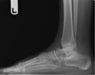
- An enlarged posterior process of the talus that can be seen on the lateral x-ray.
- Lateral x-ray taken with the foot in a pointed down position will often show the bone getting hit between the tibia and calcaneus.
- The extra small bone in the back of the ankle called the os trigonum.Muscles allowed other causes of pain in the back of the ankle including small avulsion fractures off of the tibia or fibula or fractures of the body of the talus.
MRI:
- MRI is useful to show the bone marrow swelling of the talus and/or os trigonum.
- Swelling is usually present in the back of the ankle that can be seen in T-2 weighted images.
- Helpful to have an MRI to rule out other causes of the impingement.
TREATMENT:
- Conservative treatment includes rest and the application of a boot to keep the ankle immobilized. This prevents the foot from pointing down and gives the pain in the back of the ankle an opportunity to heal. Anti-inflammatories help to reduce the inflammation. For severe pain in a fracture to the ankle, cast immobilization may be needed for 4-6 weeks. If pain is acute and moderate to severe, rest in the boot.
- If conservative treatment fails, an MRI is performed to fully evaluate the pain in the back of the ankle.
- Unresolved pain may require one or more steroid injections in the back of the ankle to Jessen some of the pain and scarring of the soft tissue that are present.
- Surgery is commonly performed on athletes and dancers due to chronic pain in the back of the ankle. For soft tissue or bony injuries, arthroscopic surgery with a posterior or lateral approach can be easily performed to remove any scar tissue and painful os trigonum if present. If an enlarged tubercle of the talus is noted this can also be debrided arthroscopically or endoscopically. Postoperatively the patients start moving the ankle within 24 hours to prevent recurrence of scar tissue and pain in the back of the ankle. A slow return to dance and sports begins at approximately 10-14 days. Full recovery can take ten or more weeks.
- Open surgical treatment via a lateral approach has also been extremely successful in our hands. A small 3-4 em incision is placed on the outside of the ankle between the fibula and . Achilles tendon. The os trigonum is easily removed. Postoperatively a very short course of immobilization of 3-4 days is done to allow the skin to heal and then early range of motion to prevent scar tissue formation is allowed. Slow return to sports and dance in as little as 10-14 days. A full return can take ten or more weeks.
- Any pathology to the flexor hallucis longus tendon is addressed via open or arthroscopic approach.
- Medial approach to the ankle is never performed in our practice due to the potential risk of injury to the nerves in the ankle and foot.
- Injuries to the flexor hallucis longus tendon are rare in isolation in the back of the ankle. It is almost always associated with a painful os trigonum. When treating the os trigonum the pain in the flexor hallucis longus tendon will usually resolve.
PROGNOSIS:
- Excellent in almost all cases when identifying the primary cause of the impingement
-----------------------------------------------------------------------------------------------------------
Symptomatic Os Trigonum
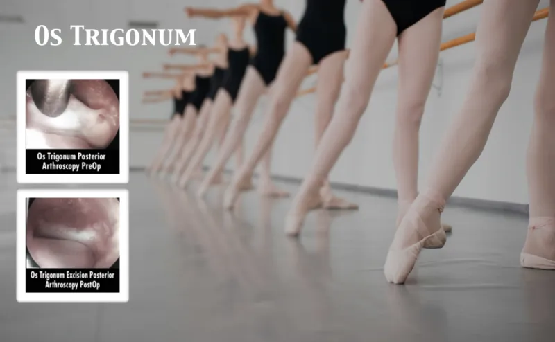
The os trigonum is a ossicle (extra bone) variant that occurs in the back of the ankle/subtalar joint. Typically occurs from birth and is present usually in both extremities. Pictured below is a female patient who had very large symptomatic ossicle that was very painful with activity especiallat any activity involved the ankle pointing the foot downward. To the left is pre-operative image, and to the right is post open resection of the ossicle.
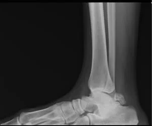
Below are bilateral x-rays of ankles in which the os trigonum can be visualized as present in both. It is the small round-like bone near the back of the ankle coming off the the back of the talus bone.
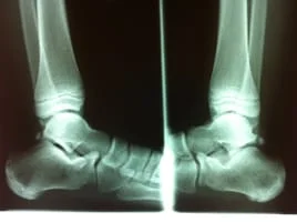
X-ray and MRI of symptomatic Os Trigonum
On MRI, the bone should be black like the other bones in this image and the bone is white indicating inflammation

MRI showing inflammation of the os trigonum with associated soft tissue swelling.

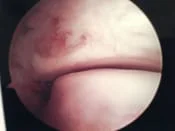
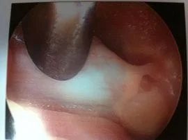
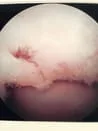
The image below is that of an intra-operative image under arthroscopy of a symptomatic os trigonum indicated by metal probe.
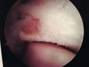
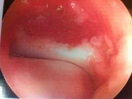
The image below is after removal of the os trigonum and now you can clearly visualize the posterior facet of the subtalar joint with healthy cartilage.
Arthroscopy pics of os trigonum attached by cartilage. It is on the left hand side.
After removal, the area can be seen free of any bone