SURGICAL PICTURES


Intraop Pic of Lateral Malleolar Avulsion fracture before and after screw fixation.


Intraop Pics of posterior anti-glide plate after ORIF fibular fracture


Intraop Pics Of Displaced Fibula Fracture After Anti-Glide Plate

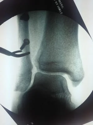
Cotton Test
This is a fibular fracture that had Deltoid rupture that causes the increased space at the inside (medial) ankle. The joint should be congruous and symmertical. After repair of the fibula, an instrument is placed around the fibula and there is traction applied laterally to check the integrity of the syndesmosis. If there is widening of the syndesmosis with lateral pull of the fibula, the test is positive for syndesmotic rupture and will necessitate stabilization of the syndesmosis.

These are pics of a patient one of our physicians saw after a gun shot wound to the ankle shattered his fibula.
The debris can be seen from the bullet

